• No products in the cart.
Checkout

BlinkTech

Author: marketing@blinktech.com.au

Application Examples – Digital Image Correlation VIC-3D

By Comments Off on Application Examples – Digital Image Correlation VIC-3D

Evaluating the Radial Force Profile of a Stent

Data courtesy of Dr. Kenneth Perry, EchoBio, LLC.


Application Overview

Medical device manufacturers have the responsibility to create and maintain efficient and safe products to keep the world population healthy. Dr. Kenneth Perry of EchoBio LLC is an industry leader and expert on the experimental validation of implantable medical devices. Recently, Dr. Perry was tasked with evaluating the radial force profile of a braided, self-expanding stent for use in correcting blocked blood vessels. Dr. Perry specifically needed to examine how the radial force changed from the end of the stent to the middle of the stent. This quantitative data is critical to the proper design and function of the stent, and Digital Image Correlation was the only technology commercially available that could provide the measurements needed at the required accuracy. Dr. Perry utilized the VIC-3D system to make such measurements with valiant success that could potentially revolutionize the medical device industry.


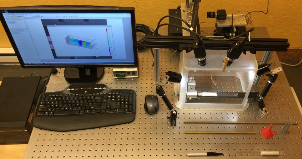
Figure 1. Experimental setup using the VIC-3D system

Dr. Perry began his experiments using perfectly straight silicone tubes that were custom made to have mechanical properties similar to a blood vessel. His experimental setup is shown above in Figure 1. The experiment involved deploying the stents into the tubes and measuring the resulting deformation. Dr. Perry knew that any simplified point or line measurement on his samples would have been misleading. This is where the VIC-3D system enters the experiment and provided Dr. Perry with the full field data he needed to be confident in his measurements and publish accurate results for his client.

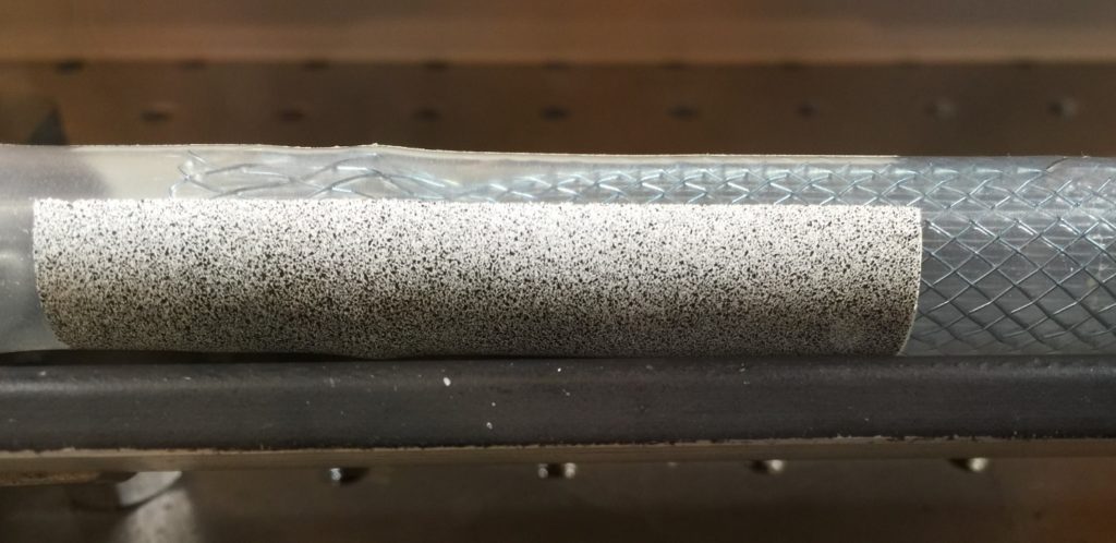
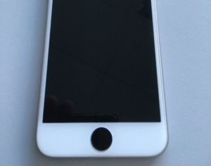
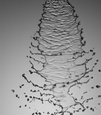
Figure 2. Speckled silicone tube with stent inserted.

The silicone tube was prepared for the experiment by having a speckle pattern applied by using off the shelf spray paint. The area of interest was masked off and a white base coat applied. The black speckles were then simply added by using an over spray technique with black spray paint. Figure 2 above shows the speckled silicone tube with the stent inserted.

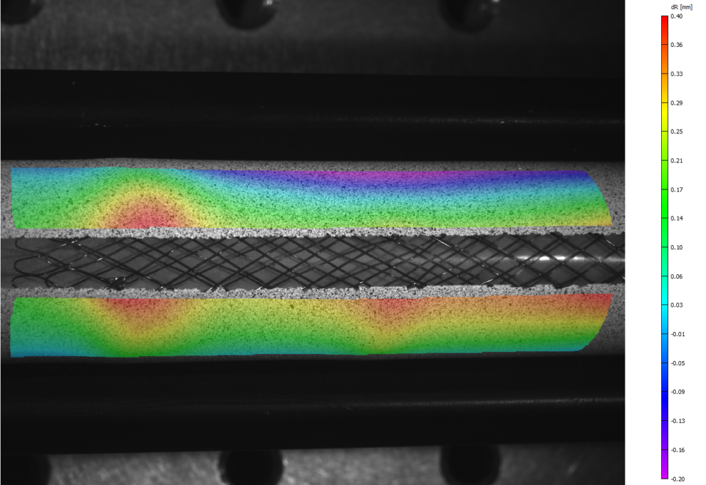
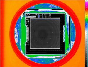
Figure 3. Change in radius contour under load from expanded stent.

The change in radius variable and resulting contour plot was the most valuable data for Dr. Perry because it clearly shows the largest force is located near the end of the stent. This contour plot, shown above in Figure 3, also indicates areas of larger radial force that could have gone unnoticed using traditional measurement techniques.

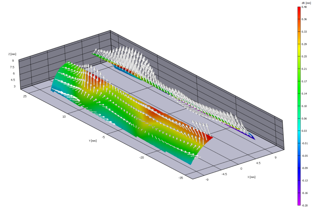
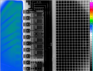
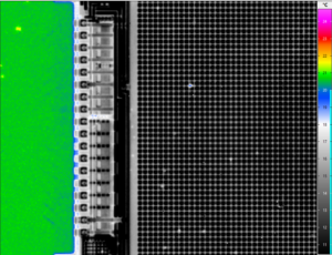
Figure 4. 3D change in radius contour plot showing displacement vectors.

Not only did Dr. Perry obtain full-field displacement data, he was also able to easily quantify the radial force profile of the stent and show the circumferential variation that resulted from the underlying woven stent structure.  Figure 4 above shows the displacement vectors of the stent in a full-field 3D contour plot. The displacement vectors show the magnitude of the resulting deformation which allows Dr. Perry to easily interpret the data as well as publish professional results for his client. The VIC-3D system gave Dr. Perry the ability to measure with confidence.

Testimonial

“Vic-3D was an amazing tool. Any simplified line or point-wise measurement would have been misleading. Vic-3D gave us full-field data we needed to be confident with the measurements.” -Dr. Kenneth Perry, ECHOBIO LLC

“We needed robust calibration, documentation and high-fidelity measurements so we designed our experiment accordingly using the VIC-3D system.”

More on VIC-3D

Thermal Imaging at Incredible Detail

By Comments Off on Thermal Imaging at Incredible Detail

We recently had a look at an old iPhone 6 using Infratec’s ImageIR 9400, one of the most versatile IR cameras on the market. The spatial resolution of these cameras mean that the experiment was able to measure down to 1.25µm/pixel.


View ImageIR

Back Illumination High Speed Imaging

By Comments Off on Back Illumination High Speed Imaging

Back illuminated imaging is a widely used imaging technique. Under the correct conditions it can provide high contrast images of object’s. This is particularly important if you are interested in an objects’ shape or size.

The idea is a simple one – a light is shone towards the camera creating a white image, the subject is then passed between the light source and the camera thus blocking the light path and creating a dark area on the image. If the subject is stationary you can focus your camera onto the objects’ profile and obtain a sharp outline. If the subject is moving, in order to freeze the motion (remove blur) and obtain a crisp outline you will need to have a sufficiently fast shutter (short exposure time).

In extreme cases a strobe light source such as a pulsed laser or LED can be used. (For more detail on lasers please see our laser range.) Using this technique you will not be able to obtain 3D information about the object shape. If you wish to resolve features on the object surface you can use a combination of front and back lighting. The technique is not limited to solids, it can equally be applied to fluids, multi-phase flows, in fact any subject that will completely or partially block the path of light to the imaging sensor.

Optical Set up: The best results are obtained with a diffuse light source. This can be achieved by passing the light through a light diffusing medium such as a ground glass or opal diffuser plate, alternatively as a low cost solution opaque drawing film works very well. Where it is not possible to position the light behind the subject you can reflect it back to the camera using a white background

Uses

  • 1.  Spray analysis
  • 2.  Droplet impaction / coalescence
  • 3.  Particle size and shape
  • 4.  Bubble formation and growth
  • 5.  Ballistics
  • 6.  Welding

Analysis of Thermal Conductivity

By Comments Off on Analysis of Thermal Conductivity

Lock-in Thermography with Infrared Camera VarioCAM® HD research 800 (exact model not available in Australia)

New materials with precisely controlled optical and thermal transport characteristics can make a large contribution to resource-saving thermal management. Scientists of the University of Bayreuth are pursuing this vision. They use infrared thermography to quantitatively determine thermal conductivity in nano- and mesostructured polymer materials.

Thermal conduction and thermal radiation are essential transport mechanisms that play a key role in various applications, from the smallest microchips to complete buildings. Their control requires a sophisticated material design that reaches into the nanometre range. Prof. Markus Retsch and his team from the Chair for Physical Chemistry 1 of the University of Bayreuth are working on the development and characterisation of such innovative materials. Modern cooling and air conditioning systems still require an external energy supply. But the cooling technology of the future should work without additional energy. To achieve this, materials are needed that selectively radiate heat. This can take place, for example, in clear weather when radiation occurs into very cold outer space through the so-called “Sky Window” in the long-wave spectral range of 8 … 13 µm, in which the atmosphere is transparent. “This process is called passive cooling,” explains Prof. Retsch, “and requires materials that emit heat via thermal radiation within a selective spectral range. At the same time as little solar energy as possible should be absorbed from the sun, for instance by improving the reflection or scattering properties of the material.”

Thin Samples Actively Excited by a Laser

On the path to such passive cooling materials, understanding of the thermal conductivity process is important. To do this, Prof. Retsch’s group is working with free-standing samples of, for example, thin polymer foils, 3D-prints, and fibre mats with a film thickness of only a few hundred micrometres. These samples are investigated with the goal of determining their direction-dependent thermal diffusivity. With this value and including the specific heat capacity and density of the sample, the corresponding thermal conductivity is calculated.

As part of the analysis, the measurement objects are excited by an intensity-modulated laser. Depending on the characteristics of the sample, the heat flux extends differently into the material (see fig. 1). The scientists actively control the entire measurement through the thermography software IRBIS® 3. The infrared camera that they use, VarioCAM® HD research 800 from InfraTec, detects the emitted infrared radiation, whose intensity varies with the lock-in modulation frequency.

Thermal Imaging Software

By Comments Off on Thermal Imaging Software

A high-quality infrared camera is the basis for successfully using infrared thermography to solve your measurement and testing tasks. However, the full potential can only be achieved with an equally powerful software. The result of Infratech’s R&D and hard work is their modular software family IRBIS® 3. Its range of functions can be configured to suit your application, so that the ideal package is available to you.

  • Modular concept for application-specific configuration
  • Compatibility with all camera models by InfraTec
  • Convenient camera control and data acquisition
  • Numerous analysis functions and tools
  • Lock-in mode for the analysis of amplitude and phase images and other complex evaluations
  • Consistent visual display of all measurement data
  • Professional compiling of thermographic reports
Find Out More

High Speed Camera Rental Australia

By Comments Off on High Speed Camera Rental Australia

A rejuvenated red Kookaburra ball has been introduced for the final two round of Austrlia’s state cricket league before the KFC Big Bash break will feature a subtly different ball which has been subject to ongoing testing and tinkering by Kookaburra in recent years.

The testing, carried out at Kookaburra’s facility in Melbourne, utilised a number of different techniques including the Photron AX200 32GB camera to film compression and impact of the ball. The camera was only needed for a short time during the testing period so a short term rental was organised along with some basic remote training and trouble shooting. The below video and outcome of the trials show just how easy the Photron PFV software and camera is to use.


Full article: https://www.cricket.com.au/news/new-kookaburra-ball-to-be-trialled-in-next-two-rounds-of-marsh-sheffield-shield-cricket-australia/2019-11-27

Promon Streamer G1 Now Available – Long Recored High Speed Camera System

By Comments Off on Promon Streamer G1 Now Available – Long Recored High Speed Camera System

The Promon Streamer G1 is the perfect long record high speed camera system for the production floor across a number of industries. The compact footprint makes the Streamer a perfect system for permanent installation on a production line. Beside the high speed recording feature and the long time recording capabilities, the built-in I/Os allow control from PLC – so it’s ready when you need it. The versatile system is available in different variations such as built-in camera, with an additional external camera, or two external cameras. The control software is based on Imaging Studio v4, all features are easy configurable by the software.

View Product

Thermal Imaging in Vet Science

By Comments Off on Thermal Imaging in Vet Science

Medical examinations of animals can be carried out in an efficient way by applying infrared camera systems. Animal’s body temperature can indicate multiple health problems. These may affect the animal itself, as for instance diseases of the udder or hoof, or they result from animal husbandry, as it is in the case of an incorrect application of saddles causing pressure points and, following, local overheating on horses. Inflammations and lameness are further problems which can be detected with the help of thermography.

The Infratec range of cameras are well equipped to handle the demands for high thermal resolution to give precise measurements that other brands are not able to provide. One such application can be seen in the research article on Evaluation of thermal pattern distributions in racehorse saddles using infrared thermography.

View Research Article

New HD Format for ImageIR® Range

By Comments Off on New HD Format for ImageIR® Range

A tremendous range of applications is typical for infrared thermography. The multitude of different tasks is synonymous with a high diversity of requirements that users have with modern infrared cameras. InfraTec meets this challenge with a completely new model of its high-end camera series ImageIR®. ImageIR® 9500 expands the company’s range of thermography systems that are focused on the global market and are suitable for tests in the mid infrared range.

High-level geometrical resolution
The special feature of the ImageIR® 9500 is clearly its cooled FPA photon detector. This is based on highly sensitive mercury cadmium telluride (MCT) and has a 16:9 HD format with (1,280 x 720) IR pixels. Due to its high native geometrical resolution, smallest structures on large-scale objects can be analysed in detail. The combination with a high-performance microscopic lens enables the display of structures of up to 1.5 μm in size. Users save valuable time by reducing the number of required single recordings while avoiding geometrical measurement errors.

Even more efficiency is achieved by using InfraTec´s unique MicroScan function. Thanks to this, the geometrical resolution can be increased to 3.7 Megapixels in full-frame. Images of this quality show the measurement objects in extremely high resolution.

Predestined for solving the most demanding measurement and testing tasks
Based on such values it is clear that the ImageIR® 9500 is ideally suited for international usage in scientific and research institutions. Other technical features of the camera underline this profile. This includes the thermal resolution of up to 0.025 K. It supports the reliable detection of very small temperature differences on measurement objects and creates the requirements for creating noise-free thermal images. IR frame rates up to 1.5 kHz in quarter-frame together with extremely short integration times of only a few microseconds provide users with the ability to analyse fast-running thermal processes.

Comparable to other infrared cameras of the ImageIR® series, this model is also extremely flexible to configure. The camera comes with latest detector linear cooler technology. Its modular design provides convenient retrofitting with components such as motorized focus, internal high-speed shutters as well as motorised filter respective aperture wheels. This means that users can easily solve their measurement tasks – no matter what field of application they are working in.

Low Light High Speed Camera Test

By Comments Off on Low Light High Speed Camera Test

The conditions were anything but perfect at AOS’s outdoor crash day for their big insurance company client. The sun was hiding behind a heavily overcast sky, sub-optimal for high speed camera recordings – but not for the M-VIT! The new sensor with 1280 x 800 and up to 4000 frames/sec offers a perfect image without motion blur even under challenging lighting conditions.

M-VIT 1280×800 @ 2000 from AOS Technologies AG on Vimeo.